From the top down, System Function:
In a healthy state of an individual without myasthenia gravis acetylcholine receptors are not degraded and neuromuscular function remains intact. The peripheral nervous system can signal to the skeletal muscles when they need to contract. When the human body moves a signal is sent from the brain, down the spinal cord, to peripheral nerves and then to the muscle fibers. These signals travel through the nervous system via neurons with mortar neurons transmitting the signal to the muscle. The muscle can then contract which allows for movement. This movement allows us to walk, run, breath, eat and perform many other functions (Australia, 2021). Therefore as a result, the weakness and paralysis of the muscular system are not present in the healthy state because both the nervous system and muscular system can effectively communicate with one another.
Cellular and Subcellular Roles:
Individuals without myasthenia gravis have receptors that are not degraded and therefore the function of their neurons remains intact. At the neuromuscular junctions, the neurons are able to provide the molecules necessary to signal to the muscles that they need to contact. When the neuron receives a signal, or impulse that the muscle needs to contract, they receive a signal at the dendrite. This impulse travels down the axon using the myelin sheath to efficiently move the signal to the axon terminal. This signal then reaches the synapse where the impulse is released to be taken up by the muscle cell (Omar, 2020).
In healthy neurons of those without myasthenia gravis, there is effective communication be the pre-synaptic terminal of one neuron to the adjacent post-synaptic neuron. When the action potential needed for a signal to occur is reached, the mitochondria in the neuron help metabolize the formation of acetylcholine through glycerophospholipid metabolism. The acetylcholine is then transported into a vesicle. Through exocytosis, the neuron is able to release the signaling molecule, acetylcholine, across the synaptic cleft to bind to receptors that signal the need for muscular contraction (Dajas-Bailador, 2004). These neurotransmitters bind to the acetylcholine receptors and can then signal the muscular contraction in the cells (Deschenes, 1994). In those without myasthenia gravis, these receptors are not degraded, and therefore the signal can be received.
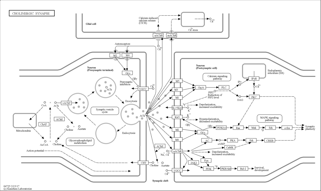
Figure 1. This image represents the process by which muscles can receive a neurological signal. The signaling pathway is visualized with acetylcholine binding to the receptors across the neuromuscular junction. The path to which acetylcholine is exocytosed from the axon (left) into the synaptic cleft is visualized. From the synaptic cleft, the acetylcholine binds to the dendrite of the next neuron (left) and some of the acetylcholine also binds to the acetylcholine receptors (top). This binding opens the ion channel of the of the receptor and signals muscular contraction (Kanehisa, 2017).
At the Bimolecular/ Atomic Level:
The released acetylcholine is able to bind to a nicotinic acetylcholine receptor. The missing acetylcholine receptors in those with myasthenia gravis prevent the acetylcholine from binding to transmit the signal to the muscle. Since the receptor is not degraded in the healthy state the receptor is then able to open the calcium ion channel in which the ions can then flow through the membrane and result in depolarization. When depolarization occurs, the muscle action potential is reached and this excitation results in muscular contraction (Ohkawara, 2021). This is the process that occurs in those without myasthenia gravis. Those with the disorder lack the receptors for the acetylcholine to bind to initiate this signal transduction cascade and as a result experience paralysis.
The acetylcholine is released from the neurons into the synapse then bind to the acetylcholine receptor at the αγ and αδ subunits (Chou, 2004). Specifically for these binding interactions to take place, glycosylation must occur at the N-terminal domain of the α subunit, and disulfide binding needs to occur. When all subunits are expressed, this forms the acetylcholine binding receptor (Green, 1998). When acetylcholine binds to the receptor, this interaction opens the ion channel of the receptor which is a pentameric ring so that the ions can flow through and allowed the muscles to contract. The ligand binding domain helps bind acetylcholine to activate the ion channel. A key residue of the receptor that is essential for the ligand of acetylcholine to bind was tyrosines (Olsen, 2014).
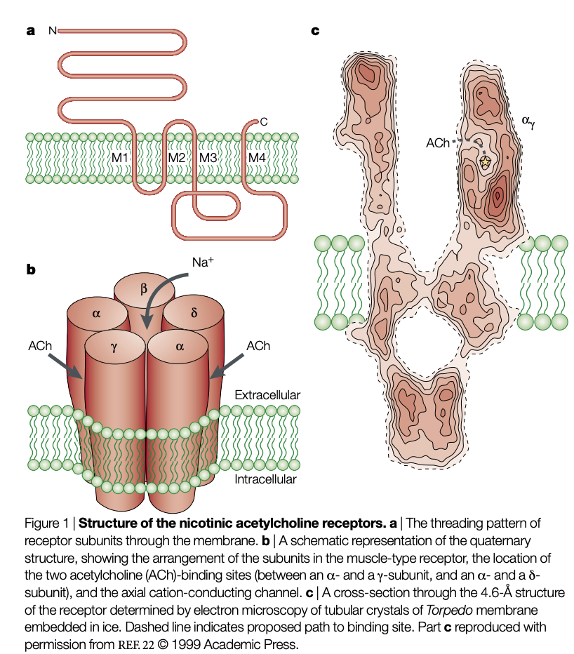
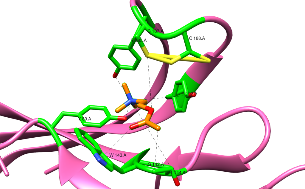
Figure 3. These figures represent the healthy state binding of acetylcholine (orange) to the nicotinic acetylcholine receptor (pink). This image represents one of the binding pockets in the pentameric ring that acetylcholine binds to the receptor. The specific residues involved in this binding pocket are C187, C188, Y192, T144, S142, W143, Y185, and Y189 (articulated green). The multiple tyrosine residues are a common characteristic of the binding pockets on the receptor. This binding opens the receptor to allow for ions to flow and signal muscular contraction (Olsen, 2014).
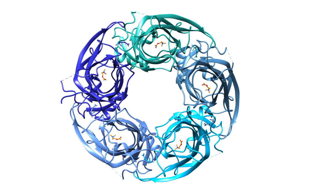
Figure 4. The right figure shows the pentameric ring of the acetylcholine receptor formed by each of its subunits (shades of blue) with acetylcholine (orange) bound throughout the ring. This binding opens the receptor so that ions may flow through the center across the membrane and muscular contraction can occur (Olsen, 2014).
Click to learn about how this process is affected in those with myasthenia gravis
The sources used:
Australia, H. (2021). Neuromuscular system. https://www.healthdirect.gov.au/neuromuscular-system
Omar, A., Marwaha, K., & Bollu, P. C. (2020). Physiology, neuromuscular junction. In StatPearls. https://www.ncbi.nlm.nih.gov/books/NBK470413/
Deschenes, M. R., Maresh, C. M., & Kraemer, W. J. (1994). The neuromuscular junction: Structure, function, and its role in the excitation of muscle. The Journal of Strength & Conditioning Research, 8(2), 103-109. https://journals.lww.com/nsca-jscr/Abstract/1994/05000/The_Neuromuscular_Junction__Structure,_Function,.8.aspx#:~:text=The%20neuromuscular%20junction%20plays%20a,in%20a%20muscle%20fiber%20action.
Green, W. N., & Wanamaker, C. P. (1998). Formation of the nicotinic acetylcholine receptor binding sites. Journal of Neuroscience, 18(15), 5555-5564. https://www.ncbi.nlm.nih.gov/pmc/articles/PMC6793068/#:~:text=Nicotinic%20acetylcholine%20receptors%20(AChRs)%20are,with%20%CE%B3%20and%20%CE%B4%20subunits.
Chou, K. C. (2004). Insights from modelling the 3D structure of the extracellular domain of α7 nicotinic acetylcholine receptor. Biochemical and biophysical research communications, 319(2), 433-438. https://www.sciencedirect.com/science/article/pii/S0006291X04009684
Brejc, K., van Dijk, W. J., Klaassen, R. V., Schuurmans, M., van Der Oost, J., Smit, A. B., & Sixma, T. K. (2001). Crystal structure of an ACh-binding protein reveals the ligand-binding domain of nicotinic receptors. Nature, 411(6835), 269-276. https://www.nature.com/articles/35077011
Dajas-Bailador, F., & Wonnacott, S. (2004). Nicotinic acetylcholine receptors and the regulation of neuronal signalling. Trends in pharmacological sciences, 25(6), 317-324. https://pubmed.ncbi.nlm.nih.gov/15165747/
Karlin, A. (2002). Emerging structure of the nicotinic acetylcholine receptors. Nature Reviews Neuroscience, 3(2), 102-114.

Hi Lauren – you picked a really interesting topic for your blog! You mention above that degradation of the acetylcholine receptors across cells prevents muscles from contracting resulting in paralysis and weakness in patients with MG. Which muscles are mainly affected and why are some affected first/more than others?
Hi Alyssa, thank you very much! In patients with myasthenia gravis, their skeletal muscles are the ones that are mainly affected by receptor degradation. This is because the skeletal muscles mainly involve voluntary contraction compared to the involuntary contraction of smooth muscle. The acetylcholine receptors are involved in the process of excitation-contraction coupling for their contraction mechanism. This process increases the intracellular calcium ion concentration necessary for contraction to occur via acetylcholine receptors. Smooth muscle utilizes a secondary mechanism to increase its intracellular calcium concentration. Smooth muscle accomplishes this by using inositol triphosphate to bind to the inositol triphosphate receptor to signal for the release of calcium. This means the smooth muscle can still release the calcium needed for contraction in the absence of acetylcholine receptors so they are not affected in those with myasthenia gravis. Skeletal muscles are affected since the degradation prevents these muscles from receiving the calcium needed to contract which is why the paralysis is a symptom of myasthenia gravis patients.
Hi Lauren,
Great topic! I often work up and have diagnosed patients with this syndrome. One of the challenges of myasthenia gravis is that testing is often not very sensitive (in fact, sero-negative myasthenia is common). Can you discuss how the different antibodies interact with the acetylcholine receptor? The most commonly tested ones are anti acetylcholine receptor modulating antibodies, anti acetylcholine receptor blocking antibodies and anti acetylcholine receptor binding antibodies. There are also two newer antibodies we test for – anti muscle specific kinase (Musk) antibody and anti LRP4 (LDL receptor related protein 4).
Thank you!