Neurological and Muscular System Dysfunction:
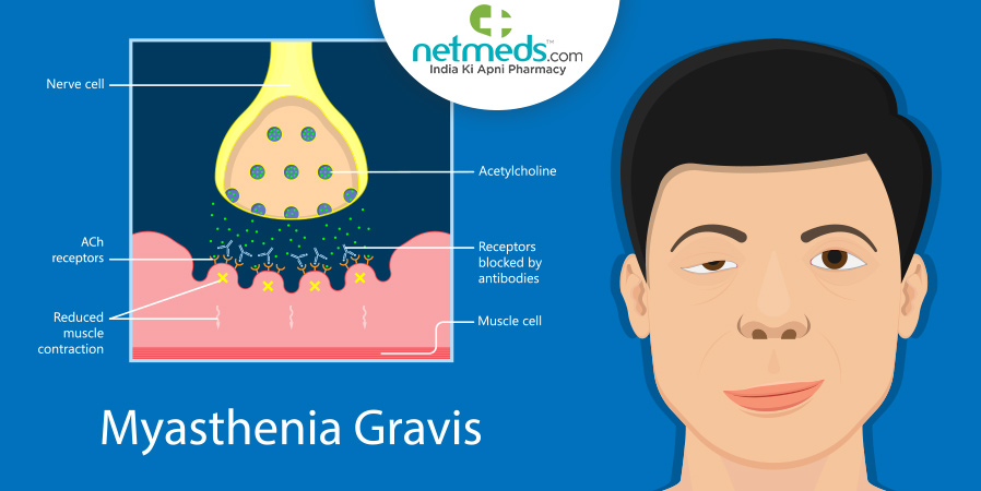
In individuals with myasthenia gravis, their muscular system and immune system are affected. The immune response attacks the nicotinic acetylcholine receptor which affects neuromuscular function. The lack of connectivity from the receptor deficiency in the nervous system prevents proper muscular contraction which leads to weakness and paralysis (Warrier, 1967). The lymphatic system was also found to be affected in those with myasthenia gravis due to the lymphocyte accumulation in those with the disorder (Buzzard, 1906). Specifically, in the lymphatic system, the thymus is affected in those with the disorder and often will develop a tumor (Bell, 1917). The thymus produces the immune cells that malfunction in those with myasthenia gravis (Norris 1936). In individuals with myasthenia gravis, the antibodies that attack the receptors come from the thymus and in 10% of patients, they have a tumor on their thymus (Ha, 2015). This results in the degradation of acetylcholine receptors which prevents proper muscle function and this presents as the paralysis and weakness seen in those with the disorder (Churchill-Davidson 1953).
The Immune Response On Neurons:
Lymphocytes are affected in those with myasthenia gravis and these immune cells were found to accumulate in the muscles of those with myasthenia gravis (Buzzard, 1906). This is because myasthenia gravis is an autoimmune disease in which autoantibodies bind to and signal the degradation of the acetylcholine receptors at the neuromuscular junction. This prevents the receptor from opening to let ions in. Therefore the muscle cells do not depolarize and will not contract (Patrick, 1973). The B cells with antibodies myasthenic IgG and Fab were found to partake in this process (Drachman, 1978). Also, with the immune response epitopes of the acetylcholine peptides on muscle cells are recognized by CD4+ lymphocytes which also contributes to the degradation of the receptors and pathogenesis of myasthenia gravis (Moiola, 1994). This degradation of receptors across the cells results prevents depolarization that prevents the muscles from contracting. This results in paralysis and weakness in the muscular-skeletal system of those with myasthenia gravis. A common case among those with the disorder is that they often do worse with physical exertion (O’Connor, 2020). This fits with the cellular response because the degradation of receptors limits the amount of physical activity one can partake in.
Pathways Effected:
Multiple pathways are affected in those with myasthenia gravis. The neuroactive ligand-receptor interaction with the nicotinic acetylcholine receptor alpha-1 malfunctions and the channel cannot uptake the acetylcholine needed (Engel, 1996). In N-Glycan biosynthesis, ALG7, ALG14, and ALG2 are all affected in the pathway of those with myasthenia gravis (Lowe, 2003). These enzymes and signaling are disrupted. When a cholinergic synapse occurs the signaling is affected in the synaptic vesicle cycle where ChAT and vAChT prevent acetylcholine from being transported into the vesicles so that it may cross the synapses of neurons. These pathways within the cell are all involved in neuromuscular signaling. Without the proper diffusion of acetylcholine across the synapse, muscles cannot be signaled to contract. This results in paralysis seen in patients with myasthenia gravis. When this paralysis occurs in the diaphragm, respiratory distress results (Bauché, 2016).
Receptor Degradation:
All cases of myasthenia gravis involve an autoimmune response in which the pathogenesis of the immune response result in the degradation of the acetylcholine receptor. Some patients have a genetic mutation in their nAchR that makes them more likely to have the autoimmune response that results in myasthenia gravis (Nichols, 2001).This protein is then degraded by an immune response that then develops myasthenia gravis and muscular weakness in patients. In the diseased state mutations in the ε-subunit of the receptor prevent proper transcription. The point mutation in the promoter of the subunit from cytosine to thymine occurs resulting in the development of myasthenia gravis. This point mutation triggers an immune response that results (Nichols, 2001). Also, single nucleotide polymorphisms in the CTLA-4 gene lead to the development of myasthenia gravis. This is a T lymphocyte protein, the promoter and first exon of the gene had certain polymorphisms that lead to thymoma. When this occurs at the +49 base pair myasthenia gravis has been shown to develop (Wang, 2002). The immune response, as a result, degrades these acetylcholine receptors and as a result, there are too few of them. The receptors were degraded because when crosslinked, the endocytosis of the receptors could occur at a faster rate. This immune response was also shown to block the receptor binding sites and prevent the ion channel from opening. In addition, those who develop myasthenia gravis have the formation of membrane attack complexes. The complex damages the muscle endplate (Ha, 2015). This prevents acetylcholine from traveling from one neuron to the next across the synapse. In the post-synaptic region, there is supposed to be an abundance of C9 at the motor endplate. When lacking structural integrity from deficient receptors there ends up being an increase in space between the junctions (Sahashi,1980).
Atomic Level Dysfunction:
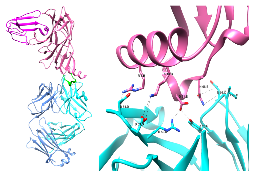
The nicotinic acetylcholine receptor is the target of autoantibodies when one has myasthenia gravis. The 3D structure of the receptor highlights the binding of the antibodies, specifically Fab, to the alpha 1 subunit of the receptor resulting in myasthenia gravis development. Specifically, this binding occurs at the main immunogenic region which spans from peptides 67-76. The essential amino acids in this binding are Asn68, Asp,71, Arg6, and Lys10, and form salt bridges as well as hydrogen bonding. This binding interaction triggers the degradation of the receptors (Noridomi, 2017). As a result, the acetylcholine necessary for neurons to signal muscular contraction cannot be diffused from synapses to move across neurons (Patrick, 1973).
Click to learn about myasthenia gravis treatment
Sources Used:
Warrier, C. B., & Pillai, T. D. (1967). Familial myasthenia gravis. British medical journal, 3(5569), 839–840. https://doi.org/10.1136/bmj.3.5569.839
https://www.ncbi.nlm.nih.gov/pmc/articles/PMC1842611/
BUZZARD, E. F. (1906). The clinical history and post-mortem examination of five cases of myasthenia gravis. Brain, 28(3-4), 438-483.
Bell, E. T. (1917). Tumors of the thymus in myasthenia gravis. The Journal of Nervous and Mental Disease, 45(2), 130-143.
Norris, E. H. (1936). The thymoma and thymic hyperplasia in myasthenia gravis with observations on the general pathology. The American Journal of Cancer, 27(3), 421-433.
Churchill-Davidson, H. C., & Richardson, A. T. (1953). Neuromuscular transmission in myasthenia gravis. The Journal of Physiology, 122(2), 252. https://www.ncbi.nlm.nih.gov/pmc/articles/PMC1366112/
Patrick, J., & Lindstrom, J. (1973). Autoimmune response to acetylcholine receptor. Science, 180(4088), 871-872.
Moiola, L., Karachunski, P., Protti, M. P., Howard, J. F., & Conti-Tronconi, B. M. (1994). Epitopes on the beta subunit of human muscle acetylcholine receptor recognized by CD4+ cells of myasthenia gravis patients and healthy subjects. The Journal of clinical investigation, 93(3), 1020-1028.
https://www.ncbi.nlm.nih.gov/pmc/articles/PMC294026/
Drachman, D. B., Angus, C. W., Adams, R. N., Michelson, J. D., & Hoffman, G. J. (1978). Myasthenic antibodies cross-link acetylcholine receptors to accelerate degradation. New England Journal of Medicine, 298(20), 1116-1122.
O’Connor, L., Westerberg, E., & Punga, A. R. (2020). Myasthenia gravis and physical exercise: a novel paradigm. Frontiers in Neurology, 11, 675.
https://www.ncbi.nlm.nih.gov/pmc/articles/PMC7403401/
Engel, A. G., Ohno, K., Milone, M., Wang, H. L., Nakano, S., Bouzat, C., … & Sine, S. M. (1996). New mutations in acetylcholine receptor subunit genes reveal heterogeneity in the slow-channel congenital myasthenic syndrome. Human molecular genetics, 5(9), 1217-1227.
https://pubmed.ncbi.nlm.nih.gov/8872460/
Bauché, S., O’regan, S., Azuma, Y., Laffargue, F., McMacken, G., Sternberg, D., … & Nicole, S. (2016). Impaired presynaptic high-affinity choline transporter causes a congenital myasthenic syndrome with episodic apnea. The American Journal of Human Genetics, 99(3), 753-761.
https://pubmed.ncbi.nlm.nih.gov/27569547/
Lowe, J. B., & Marth, J. D. (2003). A genetic approach to mammalian glycan function. Annual review of biochemistry, 72(1), 643-691.
https://www.annualreviews.org/doi/abs/10.1146/annurev.biochem.72.121801.161809
https://www.kegg.jp/entry/H00770
Nichols, P., Croxen, R., Vincent, A., Rutter, R., Hutchinson, M., Newsom‐Davis, J., & Beeson, D. (1999). Mutation of the acetylcholine receptor ε‐subunit promoter in congenital myasthenic syndrome. Annals of Neurology: Official Journal of the American Neurological Association and the Child Neurology Society, 45(4), 439-443.
Wang, X. B., Kakoulidou, M., Qiu, Q., Giscombe, R., Huang, D., Pirskanen, R., & Lefvert, A. K. (2002). CDS1 and promoter single nucleotide polymorphisms of the CTLA-4 gene in human myasthenia gravis. Genes & Immunity, 3(1), 46-49. https://www.nature.com/articles/6363816
Sahashi, K., Engel, A. G., Lambert, E. H., & Howard Jr, F. M. (1980). Ultrastructural localization of the terminal and lytic ninth complement component (C9) at the motor end-plate in myasthenia gravis. Journal of Neuropathology & Experimental Neurology, 39(2), 160-172.
https://academic.oup.com/jnen/article/39/2/160/2613146?login=true
Noridomi, K., Watanabe, G., Hansen, M. N., Han, G. W., & Chen, L. (2017). Structural insights into the molecular mechanisms of myasthenia gravis and their therapeutic implications. Elife, 6, e23043.
https://elifesciences.org/articles/23043
Patrick, J., & Lindstrom, J. (1973). Autoimmune response to acetylcholine receptor. Science, 180(4088), 871-872.
https://www.science.org/doi/epdf/10.1126/science.180.4088.871?adobe_mc=MCMID%3D50757547858844446241803403673976370587%7CMCORGID%3D242B6472541199F70A4C98A6%2540AdobeOrg%7CTS%3D1679322271
Ha, J. C., & Richman, D. P. (2015). Myasthenia gravis and related disorders: Pathology and molecular pathogenesis. Biochimica et Biophysica Acta (BBA)-Molecular Basis of Disease, 1852(4), 651-657. https://www.sciencedirect.com/science/article/pii/S0925443914003640

A helpful tool in the diagnosis of myasthenia gravis is electromyography (EMG)? Given your knowledge of the pathophysiology of this disease, what might the EMG show in a patient with myasthenia gravis, and why might this be the case?
Zachary Shuler, M.D.
Muhlenberg College Class of 2015
Hello, in patients with myasthenia gravis, their EMG results will show a decrease in muscle potential from the beginning to the end of the test. Since an EMG is used to measure how well the nerve can signal to a muscle, a decrease in the signal over time would present in an individual with myasthenia gravis. This decrease in the action potential of a muscle is representative of the decreased number of acetylcholine receptors in a patient with myasthenia gravis. The limited receptors decrease the depolarization by calcium ions for the muscle to contract. Therefore over time, the muscle action potential weaken as less acetylcholine is able to bind to the receptor and signal the action potential.
Hi Lauren, I thought your review was very thorough and educational! I’m very interested in the genetic form of this disease where the N-box of the promoter region is mutated. It seems like there is a significant reduction in the transcription of the CHRNE gene, and in more severe cases even results in the lack of production of this subunit all together. Since the full form of the protein is a pentamer, how does the lack of the Epsilon subunit impact the assembly of the nACh receptor? Does the receptor revert to the developmental form utilizing the gamma subunit or is there another way to overcome this mutation because a non-function muscular nAChR would seemingly result in death.
Hey Lauren, hope you’re doing well. Great job on the website!
1. Since MG is an autoimmune disease that, as you proposed, occurs as a consequence of misregulation within the thymus, does MG develop when the thymus is most active (in-utero)? If so, have there been efforts to test fetuses for MG during pregnancy? Is MG linked to other autoimmune diseases?
2. Is MG similar to type 1 diabetes where an unknown trigger leads to the development of the disease? Has there been any consensus on the environmental triggers of MG?
3. T-cell development in the thymus is annoying and convoluted, but it’s worth discussing with autoimmune diseases arising from the thymus! How do the molecular pathways you propose for the development of MG tie into “normal” T-cell development in the thymus?
Thanks!
Hi, thank you very much, hope you are doing well too! To answer your questions, MG pathogenesis is found to begin in adulthood. Even though it is rare there have been cases reported of congenital myasthenia gravis. These cases result from a hereditary mutation in the CHRNE gene. This gene helps synthesize the epsilon subunit of the acetylcholine receptor. Currently, physicians do not tend to test for myasthenia in-utero, but if it is suspected the child has MG, a blood test will be done to test for antibodies of the acetylcholine receptor. MG has been found to increase susceptibility to other autoimmune diseases such as Lupus, thyroiditis multiple sclerosis, and ulcerative colitis. To answer your second question yes, due to the pathogenesis of the autoimmune disorder arising in different genders at many different stages of life, researchers have had trouble finding triggers for the disease. Research has found that cases can arise after infection, or an adverse effect of medication, but have not been able to well define what triggers the disease. The hope is that with the advancing medical technology and increased understanding of the immune system, what triggers MG can be better defined in the future. In individuals with diagnosed MG, there is a consensus that air pollutants, heat, and humidity have been shown to make MG symptoms and flare-ups worse. In patients with MG, the negative selection of T-cells in the thymus is affected and as a result, the antibodies that bind to the acetylcholine receptor are released. The specific lymphocytes released are the CD4 T lymphocytes, which in a regular immune response they stimulate the macrophages and other B cells needed. This process still occurs in those with MG but instead of signaling for a pathogen to be degraded, they signal for the acetylcholine receptor to be degraded.